

(Track record and methodological references: Core Lab Publications)
Plaque and Stenosis Imaging
Over the past decade, cardiac Computed Tomography (CT) has developed into a valuable cardiac imaging tool. CT imaging of the coronary arteries allows for non-invasive assessment of coronary atherosclerosis and narrowing of the coronary lumen. Quantitative tools are available to measure lumen dimensions as well as atherosclerotic plaque volumes. Cardiac CT can further be employed to assess coronary bypass grafts and certain types of coronary stents/scaffolds.
Non-coronary Applications and Valve Analysis
Non-coronary applications of cardiac CT include quantification of contractile ventricular function and more recently developed techniques such as myocardial perfusion and late myocardial enhancement imaging. Cardiac CT has excellent three-dimensional spatial resolution and offers unmatched structural assessment of the heart i.e., the left atrium and its appendage and the pulmonary and cardiac veins or valves.
Prior to percutaneous valve interventions, cardiac CT can display the anatomy and dimensions of the left ventricular outflow tract, which is important for procedure preparation and selection of the type and size of devices. In addition, CT can be used to assess peripheral vascular access routes.
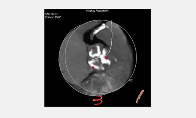
Image alludes to the assessment of calcium load in the aortic valve leaflets.
3D reconstruction of the aortic arch.
CT Syntax Score
The Syntax Score grades the complexity of coronary artery disease from a full coronary tree angiography involving the number of lesions, their functional impact, location, and complexity. The Syntax Score can be used to compare the complexity of coronary artery disease both in individual patients and entire patient cohorts. At the Cardialysis Core Lab, we also apply the newly developed multi slice computed tomography (MSCT)-derived Syntax Score.
Previous and ongoing projects involve quantitative CT angiographic follow-up after percutaneous coronary interventions (PCI), monitoring of bypass graft patency and pre-TAVI evaluations. Our services include study design and CT endpoints, acquisition guidelines, web-based training and certification of CT acquisition sites, CT data handling, independent qualitative and quantitative CT readings.
Structural Heart Core Lab
The Cardialysis Structural Heart Core Lab provides the perfect partner in conducting transcatheter therapies clinical trials (e.g. TAVR, TMTT). Over the past few years thousands of imaging data from patients enrolled in several registries and clinical trials have been successfully analyzed and adjudicated by the Cardialysis Core Lab. These data are used for CE mark and FDA submissions.
Investigator Initiated Studies
The European Cardiovascular Research Institute (ECRI) offers a platform for the design and conduct of international investigator-initiated clinical trials. ECRI acts as Sponsor ensuring compliance with ICH GCP guidelines and regulatory standards. Trial activities are executed by Cardialysis alone or in collaboration with renowned research organizations world-wide.
Monitoring Services
Cardialysis offers a consolidated monitoring network across Europe. Our clinical research associates ensure timely activation of investigational sites, high-quality data review and effective interaction with sites. With >1400 sites monitored and >200,000 patients included, Cardialysis has established a significant track record in site management.