

Magnetic Resonance Imaging
(Track record and methodological references: Core Lab Publications)
MRI Left Ventricular Analysis
Cardiac Magnetic Resonance Imaging (MRI) has become a comprehensive technology to analyze the relationships between left ventricular (LV) function, the area-at-risk and the final infarct size in the setting of acute myocardial infarction and heart failure therapies.
MRI has proven to be the most reproducible imaging modality to quantify LV function and region systolic wall thickening. Quantitative analysis is performed using semi-automatic software. Endocardial and epicardial contours are traced for the calculation of regional wall thickening using a 17-segment model that is compliant with the latest AHA guidelines.
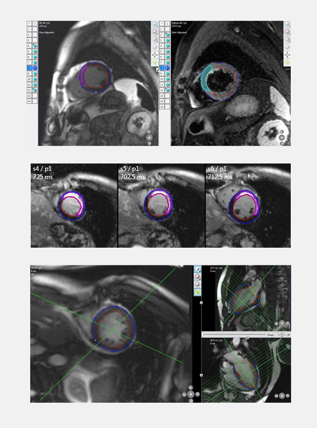
With Delayed Enhanced Magnetic Resonance Imaging (DE-MRI), an image of the myocardium can be segmented to differentiate between viable and infarcted areas of muscle tissue. The non-viable area enhances intensely while the normal myocardium maintains low signal intensity. DE-MRI is mainly applied to investigate proportional increase of viable tissue in treated acute myocardial infarction.
Our services include expert advice in protocol design and MRI endpoints, acquisition guidelines, protocol training and certification of MRI acquisition sites, MRI data handling, and independent qualitative and quantitative MRI readings.
Structural Heart Core Lab
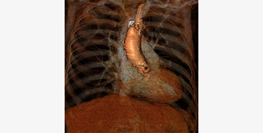
The Cardialysis Structural Heart Core Lab provides the perfect partner in conducting transcatheter therapies clinical trials (e.g. TAVR, TMTT). Over the past few years thousands of imaging data from patients enrolled in several registries and clinical trials have been successfully analyzed and adjudicated by the Cardialysis Core Lab. These data are used for CE mark and FDA submissions.
Investigator Initiated Studies
The European Cardiovascular Research Institute (ECRI) offers a platform for the design and conduct of international investigator-initiated clinical trials. ECRI acts as Sponsor ensuring compliance with ICH GCP guidelines and regulatory standards. Trial activities are executed by Cardialysis alone or in collaboration with renowned research organizations world-wide.
Monitoring Services
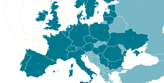
Cardialysis offers a consolidated monitoring network across Europe. Our clinical research associates ensure timely activation of investigational sites, high-quality data review and effective interaction with sites. With >1400 sites monitored and >200,000 patients included, Cardialysis has established a significant track record in site management.