

Echocardiography
(Track record and methodological references: Core Lab Publications)
The Cardialysis Echocardiography (Echo) Core Lab enables adjudication of clinically validated primary and secondary endpoints in all phases of clinical trials associated with mechanistic insights into investigational drugs and devices. Furthermore, it enables adjudication of cardiac safety for investigational compounds.
Echo Reading and Analysis
Cardialysis provides central independent Echo analysis. A team of senior technicians and cardiologists performs the analyses. This Echo team has extensive expertise in clinical study design and execution. All analyses are based on state-of-the-art recommendations and guidelines from the American Society of Echocardiography, European Association of Cardiovascular Imaging, and the Academic Research Consortium. 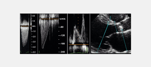
Cardialysis provides analysis of Doppler, 1D, 2D, and 3D echocardiographic modalities. Echo analysis is currently being used to investigate structural heart and heart failure. Data from a number of these studies were used for either CE marking or drug registration.
Our Echo Core Lab services are currently used in the following interventions:
- Transcatheter aortic valve replacement (TAVR)
- Transcatheter tricuspid and mitral therapies (TTMT)
- Surgical prosthetic valve replacement
- Left atrial appendage occlusion devices
- Cardiac resynchronization therapy
- Heart failure investigational compounds
The Echo Core Lab provides the following analyses:
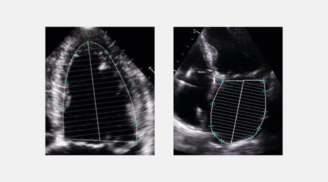
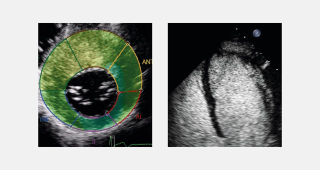
Cardiac structure and function
- 2D LV size (dimensions, areas, and volumes)
- 2D LV ejection fraction
- 2D & M-mode assessment of LV mass
- 2D LV regional wall motion
- 2D assessment of left atrial size, function
- 2D deformation imaging (strain analysis)
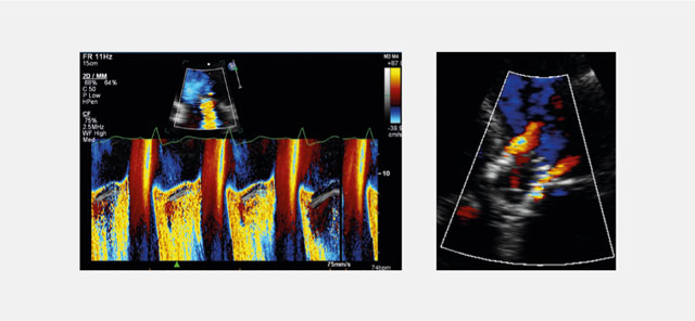
Physiologic and hemodynamic assessment
- Doppler comprehensive hemodynamic assessments
- Quantification of mitral regurgitation
- Quantification of aortic regurgitation
- Quantification of pulmonary regurgitation
- Quantification of tricuspid regurgitation
- 2D stress echocardiography and contractile reserve
- Tissue Doppler quantitative myocardial function
Together with our industry partners, we are developing vendor-independent techniques for the following Core Lab services:
- 2D speckle-tracking deformation imaging
- 3D LV volumes and ejection fraction
- 3D RV volumes and ejection fraction
- 3D aortic and mitral regurgitation
- Contrast echocardiography (perfusion/wall motion)
Structural Heart Core Lab
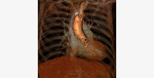
The Cardialysis Structural Heart Core Lab provides the perfect partner in conducting transcatheter therapies clinical trials (e.g. TAVR, TMTT). Over the past few years thousands of imaging data from patients enrolled in several registries and clinical trials have been successfully analyzed and adjudicated by the Cardialysis Core Lab. These data are used for CE mark and FDA submissions.
Investigator Initiated Studies
The European Cardiovascular Research Institute (ECRI) offers a platform for the design and conduct of international investigator-initiated clinical trials. ECRI acts as Sponsor ensuring compliance with ICH GCP guidelines and regulatory standards. Trial activities are executed by Cardialysis alone or in collaboration with renowned research organizations world-wide.
Monitoring Services
Cardialysis offers a consolidated monitoring network across Europe. Our clinical research associates ensure timely activation of investigational sites, high-quality data review and effective interaction with sites. With >1400 sites monitored and >200,000 patients included, Cardialysis has established a significant track record in site management.